

Man's Brain Has Air Pocket Instead Of Right Front Lobe, Doctors Shocked

Doctors treating an 84-year-old Irish man, who went to the emergency room with complaints of feeling unsteady over the past several months, were shocked to find he was missing the right frontal lobe of his brain, reports said.
The unnamed man sought medical attention after several months of unsteady walking, multiple falls and weakness on his left side. However, his medical history was clean — he didn't smoke, and rarely drank alcohol, according to a report published in BMJ Case Reports.
"There was no confusion, facial weakness, visual or speech disturbance," the doctors wrote. "He was otherwise fit and well, independent with physical activities of daily living ... and lived at home with his wife and two sons."
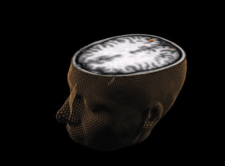
Initial tests led the doctors to believe the condition was due to old age. However, CT and MRI scans revealed to the patient’s medical team he only had a large, empty void called pneumocephalus, where the right frontal lobe of his brain should have been.
Finlay Brown, a physician who was working in the emergency department at Causeway Hospital in Coleraine, Northern Ireland, where the patient’s examination was conducted, recalled reviewing the brain-imaging scans with accompanying staff.
"(We) were all very perplexed by the images we saw!" Brown told the Washington Post in an email. "We knew immediately that there was something very abnormal. Initially, we thought perhaps the patient hadn’t disclosed having previously had some form of operation or a congenital abnormality, but … he confirmed he hadn’t."
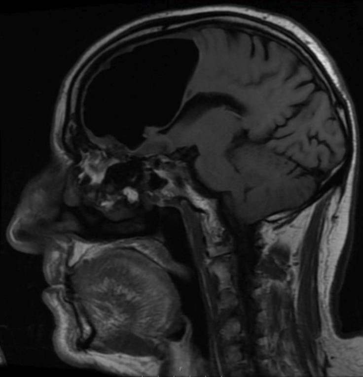
Such a condition occurs when a pocket of pressurized air forms within the cranium, which typically happens after brain surgery, the BMJ report published in February, said. In this man's case, the air cavity formed in his brain measured 3.5 inches at its longest point, which is enormous.
"In my research for writing the case report I wasn't able to find very many documented cases of a similar nature to this one," Brown, told the Washington Post.
The pneumocephalus, as discovered after the MRI, was caused by an osteoma, or a benign bone tumor that formed in the patient's sinuses and was eroding through the base of his skull.
"From speaking to the specialists, it seems it has been progressing insidiously over months to years," Brown said. "When the patient sniffed/sneezed/coughed, he would most likely be pushing small amounts of air into his head."
The report said the man's weakness on his left side was likely caused due to a small stroke, which is said to be a "rare" side effect of having an air cavity in the brain. In order to revert it, the patient had two surgeries — one to remove the tumor and the other to decompress the air pocket in the brain.
According to the BMJ report, he had been prescribed medication to prevent another stroke and was also given instructions to monitor the feeling in his left side. It added after three months, the man "remained well."
Brown and his co-authors, however, emphasized in their paper symptoms like these should always be thoroughly explored and examined.
"Because every now and then," Brown told LiveScience, "there will be a rare (or) unknown causation of these that could be overlooked."
© Copyright IBTimes 2025. All rights reserved.
- MOST POPULAR IN World


