BigBrain Atlas Provides Most Detailed 3D Model Of Human Brain Yet

Scientists have created a new three-dimensional model of the human brain with a resolution 50 times better than existing models. By taking a close look at a virtual map of gray matter, researchers may gain a better understanding of how neurological diseases arise -- and how best to treat them.
The human cerebral cortex is a marvel of intricate folds. Current models of the human brain, based on magnetic resonance imagery scans, achieve a resolution of about 1 millimeter in each direction, which sometimes loses the finer details. But the resolution of the new BigBrain model is 50 times finer, at 20 micrometers in each direction (1 micrometer is equivalent to a thousandth of a millimeter). That level of resolution allows scientists to map the detailed microstructures of the brain down to a near-cellular level.
“You can look at practically all areas in the brain,” one of BigBrain’s creators, Karl Zilles, told reporters at a teleconference with reporters on Wednesday. If you are an Alzheimer’s disease researcher, “you have the first ever brain model where you can look into details of the human hippocampus, which is the brain region extremely important for memory.”
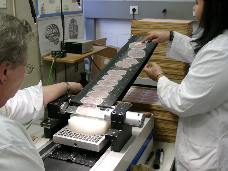
To make the model, researchers from the Research Centre Julich in Germany and McGill University in Canada used a tool called a microtome (think of it as a much more high-tech version of the machine that slices up bologna at the deli) to cut up a 65-year-old woman’s paraffin-encased brain into 7,400 ultra-thin sections.
Additional details about the brain donor’s life are not currently available. All that’s known at present is her age, sex and that she was not suffering from any known neurological diseases.
Peter Stern, one of the editors for the journal Science -- in which a paper describing the BigBrain model was published on Thursday -- said that BigBrain is bringing anatomy back to the forefront of neurological research, a field where molecular and genetic methods have held center stage for some time.
“Without really deep knowledge of the structures involved, we will never have an understanding of the data being produced by many of these other techniques and methods,” Stern said.
Researchers and members of the public alike can access the BigBrain online and view and download the brain scans and models for their own work. The potential applications are fairly wide. Lead author Katrin Amunts pointed out to reporters that one therapy for patients with Parkinson’s disease involves placing electrodes in the brain. The new brain map gives neurosurgeons a much more detailed map that could allow them to place electrodes in more precise positions. Coauthor Karl Zilles said having the anatomy of the brain at such detailed resolution could allow scientists to better determine which areas of the brain should be targeted for Alzheimer’s disease therapies.
This first high-resolution brain atlas is obviously limited. Brains do vary from person to person and change over time. Amunts said she and her colleagues plan to repeat their process in several other brains to get a wider sample.
And there is still the potential to capture an atlas at an even finer resolution -- perhaps 1 micrometer in each direction -- that would allow researchers to see even the smallest cells inside the brain. But achieving that fine a resolution would mean a lot of data, something on the order of petabytes, which most personal computers aren’t equipped to handle. Plus, going as small as one can isn’t necessarily the silver bullet to curing or treating Alzheimer’s.
“It’s important to remember that neuroscience, like all sciences, operates simultaneously at multiple scales and certain questions only make sense at certain scales,” coauthor and McGill researcher Alan Evans told reporters. “So, for instance, you wouldn’t appeal to particle physics to ask questions at the level of chemistry, but they’re both very important levels of description at the physical world.”
© Copyright IBTimes 2024. All rights reserved.


