Scientists Watch DNA Copy And Paste Itself For The First Time

Scientists have watched DNA being copied up-close for the first time, and they say their new window into the replication of genetic material shows it is more random than previously thought.
Although scientists have thought DNA replication was a more coordinated process, one that was organized so as to prevent it from becoming damaged or mutating, watching DNA copy itself in real time has given them a new take on a mechanism that is crucial to life on this planet.
Read: Making One Change to Someone’s DNA Could Have a Domino Effect
“Almost all life on Earth is based on DNA being copied, or replicated, and understanding how this process works could lead to a wide range of discoveries in biology and medicine,” UC Davis explained in a statement about the research.
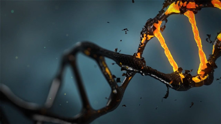
When DNA is replicated — the genetic version of copying and pasting — the two strands of its iconic double helix shape split apart like a zipper. The two separated strands are called the leading strand and the lagging strand because of the two opposite directions in which new genetic material is built on top of them. The new genetic material formed and attached to the leading strand is an exact match to what originally split away from it to become the lagging strand, and vice versa — which means when the process is complete, there will be two double-helix DNA strips that are identical to both one another and to the single strip that preceded them. This entire process is done with the help of enzymes.
Scientists had believed replicating DNA “must be coordinated to avoid the formation of significant gaps in the nascent strands,” a study in the journal Cell said.
That has to do with the different directions in which the new DNA is formed. On the leading strand, the new DNA can be built out starting from the very end where the helix started to split and moving toward the other end, toward the fork in the double helix, making for a smooth line of new genetic material. But on the lagging strand, as the name vaguely suggests, the new DNA can only be built in the opposite direction. That creates a problem because when the genetic replication begins, there is no “opposite end” yet for it to start on and meet the leading strand in the middle — there is just the fork in the double helix where the strands are still being unzippered. So the enzymes build upon the lagging strand in a rougher motion, one that is perhaps analogous to boat paddles moving on water: Just as the paddles move ahead, are dipped into the water and then move backward relative to the boat, the enzymes jump ahead on the lagging strand and build backward. When their section is done, they leap forward again to a new spot, then start building backward again until they’ve closed the gap in front of the previous section.
Read: DNA Proves Ancient Egyptians Were More Asian Than African
Although their replications move in opposite directions, the strands have been thought to be coordinated so that one strand doesn’t lead or lag by too much and potentially damage or mutate the strain. But after looking at the process more closely than ever and in real time, the researchers saw that the two strands “function independently,” the study said. They go at roughly the same rate overall, so as to complete their task at the same time, but they move along at varying speeds throughout, complete with pauses in replication. “These features imply a more dynamic, kinetically discontinuous replication process.”
The researchers were specifically watching DNA replicate in E.coli bacteria on a glass slide. Those bacteria are different from human DNA, but one thing they have in common to our species — and many others — is how their DNA multiplies. Assisting the scientists in viewing the DNA replication was a dye that would stick to a completed double helix but not to an incomplete single strand. The result was that they could watch the pace and pauses of two growing double helixes.
In the bacteria, the differences in how fast or slow the DNA replicates at any given time “can vary about tenfold,” UC Davis microbiology and molecular genetics Professor Stephen Kowalczykowski, one of the researchers, said in the university statement. “We’ve shown that there is no coordination between synthesis of the two strands. They are completely autonomous.”
And this new finding offers insight into how biochemical processes in the body work.
“It’s a real paradigm shift, and undermines a great deal of what’s in the textbooks,” he said.
© Copyright IBTimes 2024. All rights reserved.


